Please use Microsoft Edge, Google Chrome or [Firefox](https://getfirefox. com/).


ADMEDICO Augenzentrum (Olten)
Do you own or work for ADMEDICO Augenzentrum?
About Us
Philosophy
Patients should feel comfortable with us.
We at the ADMEDICO Eye Center strive to offer our patients a comprehensive service. You will be competently advised and treated by us and should feel comfortable. We want to be an understanding and helpful partner for you. Our demand for high quality services is high. For this reason, we only work with carefully selected partners.
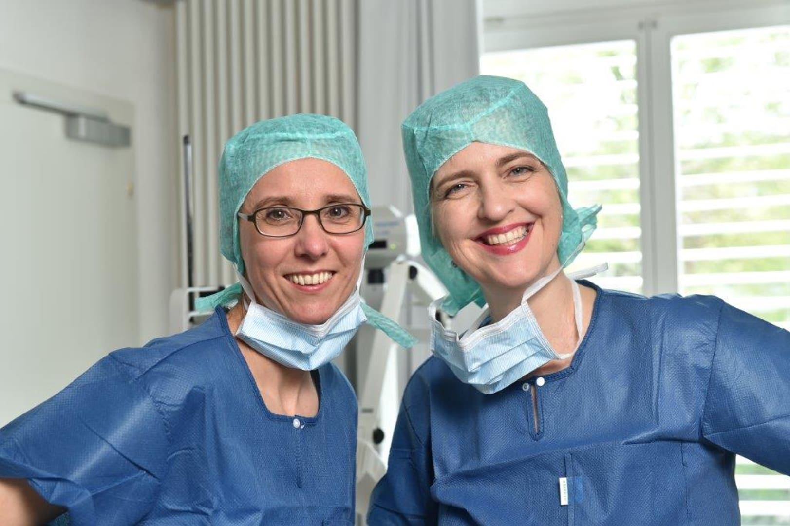
Our Team
Nicole Fichter, M.D. Specialist FMH for Ophthalmic Surgery, especially Reconstructive and Oculoplastic Surgery
Dr. med. Helga Reinshagen Specialist FMH for Ophthalmic Surgery, especially Corneal Surgery, Cataract Surgery and Dry Eye Surgery
Dr. med. Antje Hersener Specialist Ophthalmology
PD Dr. med. Alexandra Anton Specialist in Ophthalmic Surgery, Glaucoma Specialist
Tabea Karrer Orthoptist
Barbara Peter B.Sc. Optometrist FHNW
Marie Louise von Arx Nursing specialist AKP
Jacqueline Iseli Dipl. nursing specialist HF
Sabina Zaugg Dipl. consultation assistant
Michelle Wagner Consultation assistant
Karin Wihler Head of Finance and Personnel
Eye diseases (keyword phrases)
Amotio retinae or retinal detachments
Ocular herpes, eye diseases
corneal diseases, corneal inflammations
corneal transplants, corneal bank
Astigmatism or astigmatism, keratoconus
Blepharitis or inflammation of the eyelid margins
The dry eye
Diabetic retinopathy - the diabetes of the eye
Glaucoma or green star
Hyperopia or farsightedness
Cataract or cataract
Conjunctivitis or conjunctivitis, conjunctival diseases
Myopia or nearsightedness, Botox
Presbyopia or presbyopia
Strabismus or squint of the eyes
Uveitis or choroiditis
Graves' disease, endocrine orbitopathy, hyperthyroidism, Graves, EO
Eyes Olten, Eyes Solothurn, Eyes Zofingen, Eyes Oftringen, Eyes Aarburg
Symptoms of Graves' disease
Hyperthyroidism Graves' disease is a systemic disease and can manifest itself with different symptoms in different organ systems. The disease can creep into the lives of sufferers almost unnoticed. However, there can also be a seizure-like onset of the disease with acute symptoms. In addition to symptoms of hyperthyroidism, symptoms of the immune disorder also occur. However, Graves' disease can occasionally occur without symptoms of hyperthyroidism, or even with symptoms of hypothyroidism. All variants are therefore theoretically possible. Sometimes the patients get used to the symptoms, which may be mild at the beginning, and attribute them to special life circumstances or stress. If the disease is not recognized, hyperthyroidism can lead to a life-threatening condition with hormone toxicity (thyrotoxic crisis) due to massively elevated hormone levels in the blood. In addition to the signs of alteration of hormone metabolism, symptoms may occur due to the effect of antibodies in the body of the diseased person. After initiation of hyperthyroidism therapy, hypothyroidism often occurs in the further course. In order to be able to recognize the symptoms of hyperthyroidism and/or hypothyroidism as early as possible and consequently to treat them, the affected persons themselves should be aware of the symptoms and report to the doctor when they occur.
A special feature of Graves' disease are the symptoms when the eyes are involved. The cause of the eye disease (endocrine orbitopathy) is also thought to be the production of antibodies against antigens (special protein structures on the cell surface) of the eye muscles and other tissues of the eye sockets. There is a close relationship to the amount of thyroid hormone in the blood. Some people suffering from Graves' disease may also have other autoimmune diseases. It is always important to consider searching for or ruling out other autoimmune diseases in cases of unclear and confusing symptoms in Graves' disease.
For more information on Graves' disease, visit www. basedow.ch. Symptoms of hyperthyroidism Symptoms of hormone toxicity (thyrotoxic crisis) Symptoms of immune disease Symptoms of hypothyroidism Symptoms of eye disease Diabetes and thyroid disease
Corneal disease and corneal transplantation Keradonum
The cornea is the windshield of the eye and about half a millimeter with a diameter of 10 - 11 millimeters. It consists mainly of three layers: On the surface lies the outer layer, about 70 micrometers (70 hundredths of a millimeter) thick, the epithelium: it is bacteria and waterproof, constantly growing back from the edge to the center of the cornea and releasing its top cell layers into the tear fluid. It heals very well and is thus similar to the skin. Together with the tear film on top of it, it forms the optical surface of the eye, which is very important for imaging. Below it lies the middle layer (stroma) of the cornea (about 500 microns = half a millimeter), which is formed by specialized connective tissue cells. The stroma is a special tissue, because it is without blood vessels, has high mechanical strength and at the same time is optically perfectly permeable to the parts of light visible to us. It is in a dehydrated state, which is maintained only by constant pumping out of water. The inner layer, the endothelium, which is a single layer of cells covering the inner membrane of the cornea (Descemet's membrane) and adjoining the interior of the eye with the ocular fluid (aqueous humor), is responsible for the pumping and thus the transparency of the cornea. The endothelium and also the corneal stroma are nourished from the aqueous humor of the eye.
For very different reasons, the cornea can experience a deterioration of its optical properties. Some of them are briefly described here.
*Changes in the shape of the cornea
Protrusions, bulges, or distortions due to scarring interfere with imaging to such an extent that patients may have very poor visual acuity even with an otherwise clear cornea. A particular hereditary progressive protrusion is keratoconus, a cone-like taper of the cornea. In early stages, adequate correction or stabilization of the protrusion can be achieved with a contact lens or curing of the cornea through cross-linking therapy. If the protrusion is severe or the contact lens is unstable, surgical replacement of the shape-altered cornea is necessary (= corneal transplantation). The complete cornea is transplanted centrally with a diameter of approx. 8 mm.
Cloudiness and deposits in the corneal middle layer (corneal stroma).
They can be caused by metabolic diseases, which are mostly hereditary and affect only the cornea or the whole person. Scar tissue in the cornea, such as can occur after herpes infections or corneal injuries, may lead to a significant deterioration of vision that can no longer be influenced by medication. Here, a corneal transplantation with a complete graft can help.
Failure of the corneal inner layer (corneal endothelium).
Failure of the pumping function of the inner layer leads to chronic swelling of the cornea. In this condition, the tissue is no longer clear and transparent, but milky cloudy with a corresponding deterioration of the optical properties. The finest water bubbles reach under the uppermost cell layers of the cornea, where they can cause a foreign body sensation, burst painfully or even promote the penetration of bacteria into the cornea. In this disease, only the inner layer can be transplanted with the latest technology, i.e. lamellar corneal transplantation.
Dry eye
According to the definition, dry eye is based on a chronic inflammatory wetting disorder. This manifests itself with a variety of symptoms such as eye pain, burning sensation, foreign body sensation, itching, feeling of pressure or eye fatigue. The tear film, which consists of 90% water and 5% each of mucus and fat, then no longer wets the eyes continuously and sufficiently. Often the composition of the tear film is no longer balanced, and there is a lack of fat admixture. The fat comes from sebaceous glands of the eyelids, the so-called meibomian glands. If these no longer function well for various reasons, the fatty component in the tear film is missing, with the result that the aqueous component evaporates too quickly and the above-mentioned complaints occur.
In the case of dry eye, in addition to the use of tear substitutes, it is often necessary to clean the edge of the eyelid, which is easy to do, and to use anti-inflammatory eye drops from time to time. In the case of more severe forms of progression, further therapeutic components are also used
Cataract surgery
Cataract surgery is one of the most common and best standardized surgeries worldwide.
Cataract cannot be treated with medication. The principle of the operation is based on the removal of the clouded lens and its replacement with an artificial lens (intraocular lens). The necessary refractive power of this artificial lens can be precisely calculated in advance based on the length of the eyeball and the curvature of the cornea.
One eye is always operated on first. If the other eye is also affected, the second operation can be performed a few days later.
The eye is only locally anesthetized before the operation, i.e. made insensitive by eye drops. General anesthesia is necessary only in exceptional cases. During the operation, a 1.8 to 2.5 mm small incision is made at the edge of the cornea. Through this incision a tiny piece of the anterior capsule is removed. Then, using a special ultrasound device, the contents of the lens are finely crushed (phacoemulsification), removed with a suction rinsing device, and removed. What remains is the ultra-thin capsular bag, which serves as a support for the artificial lens. Next, the artificial replacement lens is inserted. It is made of an elastic transparent material that is pushed through the tiny opening and then unfolds and centers itself. The access incision is so small that it does not need to be closed by a suture, but closes like a valve.
The artificial lens remains in the eye for life, meaning it does not need to be removed or replaced later.
There are different types of lenses:
*Standard lens The cost of the standard lens is covered by health insurance. Depending on the strength, you can usually see sharply with this lens either at a distance or at close range, if necessary, without glasses. For the other distance, you will need corrective glasses.
*Aspherical lens This lens improves contrast vision, especially in low light conditions such as twilight, night or fog.
Toric lenses for astigmatism. Astigmatism can be corrected by individually calculated and manufactured artificial lenses. As a result, the perception of the environment is less distorted and blurred.
Combined near and distance lenses. In certain cases, this artificial lens enables distance and near vision that is largely free of glasses. If you are interested, a comprehensive preliminary examination will determine whether you are a suitable candidate.
Glaucoma surgery
A glaucoma is an eye disease in which the intraocular pressure is increased and/or insufficient blood flow leads to a change in the optic nerve (papilla) in the affected eye, which is typical of glaucoma. The goal of treatment of the disease is to slow down or prevent the progression of the disease by lowering the intraocular pressure.
If despite careful application of medications (drops, ointments, tablets) no sufficient reduction of intraocular pressure can be achieved, the following surgical treatment options are available, which vary in suitability depending on the form of glaucoma and the stage of the disease.
□ Laser coagulation of the meshwork (selective laser trabeculoplasty (SLT)).
After anesthetizing the cornea and placing a contact lens, low-dose laser light is applied to selected sections in the chamber angle. During the treatment, you sit with your chin propped up at a so-called laser slit lamp. This procedure is suitable for low intraocular pressure elevation or in the early stages of the disease.
□ Iridotomy with the laser.
The intense light of a laser is used to incise the iris and allow the intraocular fluid to pass directly from the posterior to the anterior chamber of the eye. The surface of the eye is anesthetized by drops so that a special contact lens can be painlessly placed on the cornea. Through this lens, a precisely targeted laser beam is directed with numerous individual "shots" to the iris, where it leads to the punctual dissolution of the tissue that has been hit. This produces a cracking sound. Since the iris is largely insensitive to pain, the iridotomy is usually painless. During the treatment, you sit with your chin propped up at a so-called laser slit lamp.
□ Sclerotherapy of the iris body (cycloplethotomy, cryocoagulation).
This method involves reducing the production of intraocular fluid by destroying part of the radiolucent body with cold (cryocoagulation) or heat (laser coagulation). This is done with the eyelids open by placing a cold or laser probe at various points on the conjunctiva. Then the cold or heat penetrates the conjunctiva and the eye wall without damaging them, causing the underlying ray body to slough off. As a result, less aqueous humor is subsequently formed.
□ Filtration surgery, trabeculectomy, goniotrepanation.
An additional drain is created by cutting out a small piece of the eye wall and creating iris windows in this area. A slight protrusion forms on the conjunctiva, which is called a filter pad. The aqueous humor can now drain out of the eye under the conjunctiva into the orbit.
□ Filtration surgery with implants (XEN, Preserflow).
The aqueous humor is drained from the eye chamber under the conjunctiva and thus into the orbit via a small implant (gelatin tube, plastic tube), thereby lowering the intraocular pressure.
□ Valve operation
A thin tube is passed from the anterior chamber of the eye through the eye wall into the tissue behind the eye. Through this tube, the intraocular fluid is drained into the eye environment.
□ Trabeculectomy ab interno (e.g. trabectome, Kahook Dual Blade).
Using an instrument, the trabecular meshwork is removed from the inside of the eye over a few hours to reduce the drainage resistance of the natural drainage pathways.
□ Ventricular Angle Implants (e.g. i-Stent)
In this procedure, small implants are inserted into the trabecular meshwork in the ventricular angle. This improves outflow into the Schlemm's canal behind it. These implants are often implanted as part of cataract surgery.
**What are the chances of success?
The success of glaucoma surgery cannot be guaranteed with any of the different surgical procedures. However, depending on the underlying disease, satisfactory and long-lasting pressure reduction occurs in most cases after glaucoma surgery. Sometimes additional eye drops are necessary to lower the eye pressure. A repetition of the surgery or the performance of another glaucoma surgery may be necessary.
Surgery cannot reverse existing damage to the optic nerve and retina. Surgery is performed only to preserve the remaining visual field and vision.
Our Partners
We work intensively with the following partners:
EUGOGO European Group on Graves Orbitopathy University Eye Hospital Basel Orbita Centrum Amsterdam Hirslanden Clinic Aarau Eye Clinic Lucerne Solothurner Spitäler AG
Kerradonum.ch
Pallas Kliniken Olten
Directions Eye Center Olten
Bus from the train station (5 minutes): Use exit Stadt and take bus line 2 (direction Trimbach) to bus stop Kantonsspital.
Taxi Cab Bur: 062 205 22 22 Aare Taxi Sigrist: 062 296 26 26
**On foot From Olten train station approx. 10 min. From Olten train station (10 minutes): Use exit Stadt, walk over the station bridge and then turn right into Amthausquai (along the Aare), after 400 meters turn left along a short forest path to the hospital (follow signs).
By car
General direction Basel. In Olten city intersection direction Trimbach.
After approx. 200m follows the Kantonsspital.
Parking in front of the building.
Languages
Location and contact
ADMEDICO Augenzentrum
-
office address
Fährweg 10 4600 Olten
-
Phone
0622... Show number 062 206 87 37 *
-
Write an e-mail
- Visit site Visit site
- * No listing required
Ratings and reviews
Do you wish to rate and review "ADMEDICO Augenzentrum"?

4 reviews from older local.ch. archives
Data from June 2019
* These texts have been automatically translated.
Other listers


ADMEDICO Augenzentrum